Retinal detachment is a serious eye condition where the retina separates from its normal position at the back of the eye. If left untreated, it can lead to permanent vision loss. Understanding the process—from diagnosis to recovery—can help you feel more prepared and confident about retinal detachment surgery.
Recognizing the Symptoms of Retinal Detachment
Early diagnosis is crucial for preserving vision. Symptoms of retinal detachment may include:
- Flashes of Light: Sudden bursts of light in your peripheral vision.
- Increase in Floaters: A noticeable rise in black spots or strings in your vision.
- Shadows or a “Curtain” Over Vision: Partial loss of your field of vision, resembling a shadow or veil.
If you notice any of these signs, contact an eye specialist immediately. Retinal detachment is a medical emergency, and prompt evaluation can prevent further vision loss.
Preparing for Surgery
Once retinal detachment is diagnosed, surgery is typically recommended as the best way to repair the retina. Your surgeon will choose the appropriate surgical method based on the severity and type of detachment:
1. Pneumatic Retinopexy: A gas bubble is injected into the eye to push the retina back into place.
2. Scleral Buckling: A silicone band is placed around the eye to gently press the retina back into position.
3. Vitrectomy: The vitreous gel is removed and replaced with a gas or oil bubble to support the retina.
Before surgery, your doctor will provide detailed instructions, including fasting guidelines and arranging transportation home post-surgery. Preparing questions in advance can help ensure you feel informed and ready.
The Surgical Process
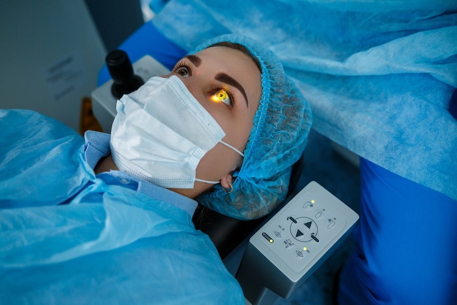
Retinal detachment surgery is often performed under local anesthesia, allowing for a quicker recovery. The procedure typically lasts one to several hours, depending on its complexity. During surgery, your eye care team focuses on ensuring your comfort and achieving the best possible outcome.
Post-Surgery Care and Recovery
Recovery from retinal detachment surgery is a critical step toward restoring vision. Your doctor will provide detailed post-operative instructions to promote healing:
- Use Prescribed Eye Drops: These helps reduce inflammation and prevent infection.
- Avoid Strenuous Activities: Rest and avoid heavy lifting or vigorous exercise.
- Follow Sleeping Position Guidelines: Depending on the procedure, you may need to sleep in a specific position to aid healing.
- Attend Follow-Up Appointments: Regular check-ins allow your doctor to monitor progress and address any concerns.
While mild discomfort, redness, and swelling are common after surgery, notify your doctor immediately if you experience increased pain or vision loss.
Long-Term Management and Vision Recovery
Although most patients can resume normal activities within a few weeks, complete healing and vision stabilization may take several months. Regular follow-up appointments are essential to monitor the retina and assess the need for additional treatments. Your doctor will guide you in managing long-term eye health and maintaining the surgical outcome.
Why Choose Retina-Vitreous Associates of Florida?
At Retina-Vitreous Associates of Florida, we specialize in providing personalized care for retinal conditions, including expert surgical management of retinal detachment. As leaders in the Tampa Bay area, our team offers:
- State-of-the-art diagnostic and surgical technology.
- A compassionate, patient-centered approach to care.
- Comprehensive support from diagnosis through recovery.
Conclusion
Facing retinal detachment surgery can be overwhelming, but understanding the process makes it more manageable. Early detection, expert surgical care, and dedicated follow-up are key to preserving vision and achieving the best possible outcome. If you’re experiencing symptoms of retinal detachment, don’t wait—schedule an evaluation with Retina-Vitreous Associates of Florida today.