Your vision feels fine. No pain, no blurriness, no warning signs.
But that is exactly the problem. The most serious eye diseases — macular degeneration, diabetic retinopathy, and glaucoma — cause zero symptoms in their early stages. By the time you notice something is wrong, the damage is often permanent.
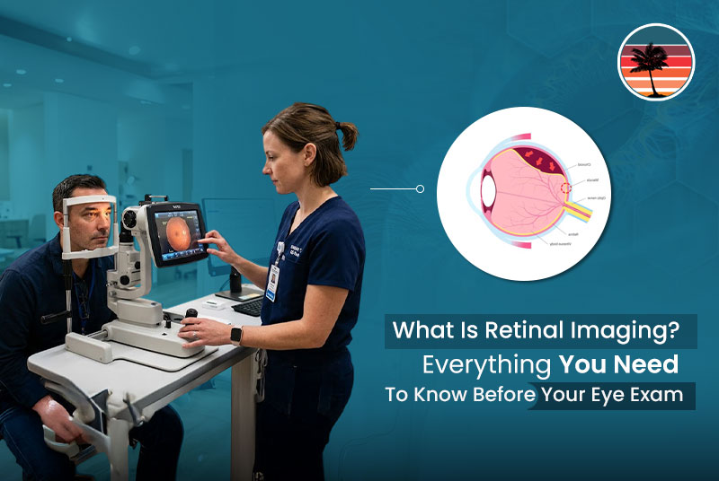
Retinal imaging changes that. It is a quick, painless test that photographs the back of your eye in high resolution — giving your retina specialist a clear view of structures no standard eye exam can reach—a brief flash of light. Nothing touches your eye. Results in seconds.
Think of it as a smoke detector for your vision. You do not wait for the fire before you check.
This guide covers everything you need to know — what the test involves, what it detects, and what to expect at your appointment.
What Retinal Imaging Shows?
At the very back of your eye lies some of the most diagnostically rich tissue anywhere in your body. This retinal scan captures it all in just a few seconds.
Your doctor will be able to identify clearly:
- Your Retina – The layer of tissue at the back of the eye that receives light.
- Your Macula – The tiny central part of the retina that controls your acute, detailed vision.
- Your Optic Nerve – The bundle of nerves that transmits visual information from your eye to your brain.
- The Vessels – The small blood vessels that run along the surface of your retina can show signs of eye disease as well as several general conditions.
Even the smallest changes in these areas will be evident on a good-quality retinal scan; that’s why the test is so useful.
Why It Matters More Than Most People Realize?
The vast majority of sight-threatening retinal diseases are silent in the early stages. You can have an incredible amount of damage developing within your eye and not know it.
Retinal imaging detects these problems before they claim your vision. Macular degeneration, diabetic retinopathy, all cause irreversible loss of sight when it is too late. Imaging provides a way to achieve a better result.
The Four Main Types of Retinal Imaging
Different types of technology examine different parts or layers of the eye. Your clinical requirements determine the appropriate technology for you.
1. Fundus Photography
This is a high-resolution color photograph of the retina, optic nerve, and blood vessels. This is the most common test used to produce permanent color baseline documentation that your doctor can compare at future visits. It is also fast and non-invasive.
2. Optical Coherence Tomography
OCT uses light waves to construct a cross-sectional scan of the retina. It provides a layer-by-layer view of your retina and has been called the “MRI of the eye”. It is the test of choice for diagnosing and managing macular degeneration, macular edema, and diabetic retinopathy.
3. Fluorescein Angiography
A small, safe amount of dye is injected into your arm. It circulates throughout your bloodstream, reaching the blood vessels in the back of your eye. Special cameras then photograph your retinal blood vessels and how they take up and drain the dye to identify abnormal, leaking, or blocked blood vessels.
4. Ultra-Wide Field Imaging
Whereas typical fundus cameras capture about 15% of the retinal surface, UWF images can capture up to 82% of the retina with a single picture. Early tears, detachments, and lesions often occur in the extreme peripheral regions of the retina, which are not usually captured by standard equipment and can therefore be missed.
What Can Be Detected With Retinal Imaging?
Apart from eyes, some systemic diseases that cause abnormal blood vessels in the back of the eye can also be identified with retinal imaging.
Eye Diseases:
- Diabetic retinopathy and diabetic macular edema
- Age-related macular degeneration (AMD)
- Glaucoma and early optic nerve damage
- Retinal detachment and retinal tears
- Retinal vein and artery occlusion
- Vitreomacular traction
- Choroidal melanoma
Systemic Diseases:
- Diabetes and its vascular complications
- High blood pressure
- High cholesterol
- Early warning signs of stroke and cardiovascular disease
Retinal imaging is therefore more than just an eye test; it is a view into your overall body health.
What Goes On During The Test?
The process from start to finish takes under 10 minutes.
- You sit at the device and place your chin on a support bar.
- Eye drops may be applied to dilate your pupils (takes about 20 minutes to take effect, but gives an extremely clear image)
- You stare into the device (left then right eye separately) at a fixed point.
- A very quick, light flash takes the picture.
- Images will be displayed immediately on the screen and discussed with your doctor.
Your eye is never touched, and the procedure causes absolutely no discomfort.
Do You Need Dilation?
Not necessarily. Many current systems (including UWF technology) can function without dilation. In more complex situations, your doctor may still recommend dilating drops.
If you do have your eyes dilated, you can expect a temporary mild light sensitivity and slight blurring of your near vision for two to four hours. It is recommended to have a driver for this purpose.
Our specialists at RVAF will tailor the imaging to your needs.
Is It Safe?
Yes. Retinal imaging is non-invasive, radiation-free, and safe for patients of all ages. The short flash of light is harmless to the eye. While fluorescein angiography involves an intravenous injection, it is well-tolerated and safe for virtually all individuals.
Who Should Get Retinal Imaging?
You would typically be offered this scan if:
- You have diabetes or pre-diabetes.
- There is a history of macular degeneration, glaucoma, or retinal disease in the family.
- You have had recent floaters, flashes of light, or a change in vision.
- You are over 40 and need a general eye test.
- You have high blood pressure or high cholesterol.
- You have a known retinal condition that requires monitoring.
Even if you have no symptoms, the retinal scan is very useful as a screening tool for many patients with early retinal problems who may not be aware of them.
Why See a Retina Specialist for This Test?
The value of your imaging depends on who is reading it. Retina Vitreous Associates of Florida utilizes fellowship-trained retinal specialists to read ALL our images. No general ophthalmologist reads your images.
RVAF has served the Tampa Bay community for over 54 years. We specialize exclusively in retinal and vitreous diseases. This expertise and focus on the particular nature of retina and vitreous diseases result in a diagnostic capability that standard general ophthalmology practices cannot match.
We are also the largest clinical trial site in the Tampa Bay region for retina and vitreous diseases. Our patients benefit not only from the latest FDA-approved medications but also, at times, breakthrough therapies in development.
Should your images reveal an issue that requires next steps, you’re already in the best hands to take them. Diagnosis, Medical Treatment, and Surgery all at the same practice.
Are you ready for your retinal imaging study?
Call RVAF to schedule a retinal imaging exam at the nearest Tampa Bay location.