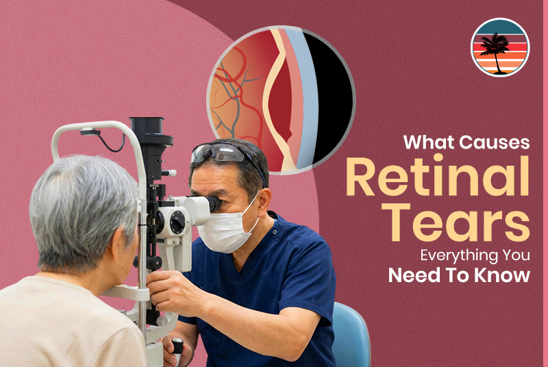
The main reason for retinal tears is a posterior vitreous detachment (PVD), where the gel-like substance in the vitreous separates from the retina. Other factors that contribute to retinal tears include aging, ocular trauma, ocular surgery, and high myopia.
A retinal tear is a medical emergency. If left untreated, it can lead to permanent vision loss. Although retinal tears can occur in people of all ages, they are most common in those over 50. Knowing the causes and risk factors will help you protect your vision.
This guide provides you with all the information you need about the causes of retinal tears, who is at risk, and when to seek immediate care.
What Are Retinal Tears?
Before we proceed to the causes, let’s first explore what happens when your retina tears.
Understanding Your Retina
Your retina is the light-sensitive tissue that lines the back of your eye. It receives light and sends signals to your brain, allowing you to see. This sensitive tissue is the inner lining at the back of your eye. Although important, it is easily damaged.
What Is a Retinal Tear?
A retinal tear is a small hole or tear in the retinal tissue. It is also known as a hole or tear. This is a serious condition that can cause vision loss. If the tear is left untreated, it can lead to retinal detachment, which may cause blurred vision, distortion, and vision loss in the affected area.
Why Retinal Tears Are Important?
Retinal tears are the most common cause of retinal detachment. If left untreated, retinal tears can cause blindness.
The retina can be thought of as the “vision keeper” that needs to be protected. When the retina tears, quick action can prevent further vision loss.
The Primary Cause: Posterior Vitreous Detachment (PVD)
Most retinal tears occur as a result of the normal aging process of the eye.
What is PVD?
PVD is the acronym for the separation of the vitreous gel from the retina. The vitreous is the clear gel that fills the eye. At birth, the vitreous is firmly attached to the retina, but as one ages, it liquefies and shrinks.
How PVD Causes Tears?
In most cases, the vitreous separates without any problems. However, for some individuals, the vitreous becomes “sticky” and pulls more strongly on the retina. As a result of the stronger pull on areas where the vitreous is strongly adherent, a tear in the retina may develop. Tears most commonly occur spontaneously in PVD at the sites where the vitreous is strongly adherent.
PVD Statistics
PVD is a normal aging process that will eventually occur in most people. However, between 8% and 22% of people with acute symptomatic PVD will go on to develop retinal tears. These tears typically occur in the superotemporal quadrant of the retina, the upper outer part, and are most prevalent in individuals over 50.
The Process Explained
As we grow older, changes occur in the gel that fills the eye. This gel is called the vitreous. It can shrink, thicken, or liquefy. This leads to its separation from the retina. In most cases, this separation takes place smoothly.
In some people, the vitreous is naturally stickier. When it separates, it may cause unusual traction. The area with the strongest adhesions may experience a tractional force. This can lead to retinal tissue tearing.
Consider the process of removing tape from paper. Sometimes, it comes off smoothly. Other times, it tears the paper.
Why Some People Experience Tears and Others Don’t?
The strength of adhesion and the consistency of the vitreous can vary from individual to individual. Moreover, the retina’s susceptibility can vary depending on its thickness and health.
The rate of PVD is also essential, as sudden retinal detachment increases the risk of retinal tears. Just as fabric wears unevenly, some areas remain strong while others fail.
Secondary Causes of Retinal Tears
While PVD is the main reason for most cases of retinal tears, other conditions may also affect your retina.
1. Eye Trauma and Injury
A physical blow to the eye or head can cause retinal tears. These include car accidents, sports injuries, falls, and physical violence.
A substantial impact can cause bruising, scarring, or a retinal tear. Trauma poses both immediate and potential risks. Retinal scarring can increase its vulnerability to future retinal tears.
2. Eye Surgery Complications
The risk of retinal tears is highest after cataract surgery, though other eye surgeries may also increase it. Risk factors after surgery include less mature cataracts before surgery, any complications that may arise during surgery, and larger axial lengths in highly myopic patients.
Tears can occur during surgery or in the postoperative period. Using meticulous surgical technique and proper postoperative care can help prevent tears.
3. Severe Nearsightedness (High Myopia)
High myopia leads to elongation of the eyeball, stretching the retina. This increased stretch makes the retina more susceptible to damage and, more importantly, tears.
The reason is simple: the thinner, stretched retina is more prone to tearing due to the tension caused by the elongated eyeball. Myopia significantly increases the risk of tears.
4. Retinal Thinning and Degeneration
Lattice degeneration is a fragile area of the retina, a pre-existing condition that affects 7-8% of the population. This pre-existing condition makes the retina fragile.
As we age or develop a disease, degeneration can further weaken the area, making an already thin spot more susceptible to a tear.
5. Aging-Related Changes
Natural weakening occurs as retinal tissue ages. The years of wear begin to take their toll. Aging can occur alongside other risk factors, thereby increasing overall risk.
Complete Risk Factor Checklist
Knowing your risk factors enables you to protect your vision proactively.
1. Age-Related Risk Factors
The primary risk factor is advanced age, particularly after age 50. The risk of developing PVD increases with age, and as time passes, the risk of multiple risk factors also accumulates.
2. Medical and Eye Health History
A history of retinal tear or retinal detachment is a significant risk factor. If the other eye gets a tear or retinal detachment, it is a high-risk indicator. A family history of retinal problems indicates genetic susceptibility.
Lattice degeneration is the development of thin areas in the retina. Retinal degeneration is the weakening of the retinal tissue. Both of these conditions are major risk factors for a tear.
3. Vision and Refractive Conditions
High myopia, also known as severe nearsightedness, is a significant risk factor. The longer the eye’s axial length, the greater the retinal stretching, the greater the myopia, and the higher the risk.
4. Systemic Health Conditions
Diabetes and diabetic retinopathy affect the retina, while sickle cell disease can impair blood flow. Inflammatory conditions damage tissues, and autoimmune conditions can affect the tissues of your eyes. Certain cancers can also increase the risk.
5. Surgical and Trauma History
A history of eye surgery, especially cataract surgery, can make the eye more susceptible to complications. Trauma, with its typical injuries, can also increase the risk. The absence of the natural lens (aphakia) increases the risk, and even with an artificial lens (pseudophakia), the risk is slightly higher.
6. Developmental and Congenital Factors
Retinopathy of prematurity affects the lives of babies who are born prematurely, and this is further exacerbated by the congenital retinal deficiencies that are also present from birth.
7. Medication-Related Risks
Pilocarpine, a glaucoma drug, constricts the pupils. Other drugs that cause small pupils may also increase the risk.
8. Lifestyle and Environmental Factors
Playing contact sports increases your chances of injury. Eye injuries in occupational settings are an essential consideration. Without protective eyewear during risky activities, you are at risk.
Risk Assessment Table
| Risk Category | Examples | Risk Level |
| Age | Over 50 | High |
| Personal History | Previous tear | Very High |
| Family History | Parent with detachment | Moderate–High |
| Eye Conditions | High myopia, lattice degeneration | High |
| Systemic Disease | Diabetes, sickle cell | Moderate |
| Surgery | Recent cataract surgery | Moderate |
| Trauma | Contact sports | Variable |
RVAF’s Expertise in Retinal Tear Diagnosis and Treatment
Trauma, surgical complications, and high myopia are the most common causes of retinal tears. By understanding your risk factors, you can remain vigilant. Learn the symptoms and seek immediate care if you experience them.
Your vigilance each day will serve you well in the years to come. Contact Retina-Vitreous Associates of Florida for your retinal needs and for emergency care. We have diagnosed and treated thousands of retinal tears and have been the innovators of retinal care in the Tampa Bay region.
At each office visit, we offer a comprehensive dilated examination, combined with state-of-the-art retinal imaging, providing a detailed retinal evaluation. We develop a personalized risk assessment based on your individual risk factors, and, when indicated, we promptly treat you. For high-risk patients, we provide ongoing observation and, most importantly, caring, expert attention throughout the patient’s experience.
If you are experiencing flashes of light, floaters, or vision changes, call RVAF today for an emergency evaluation.