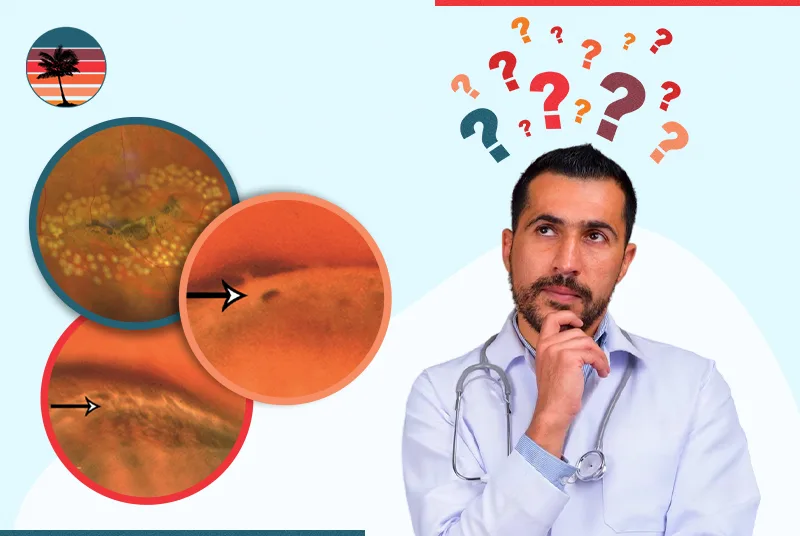
Lattice degeneration is a relatively common retinal condition in about 6–10% of the general population. It results in thinning of the peripheral retina and is a risk factor for retinal holes, atrophic holes, and, in some instances, retinal tears and retinal detachment.
Although this condition is usually asymptomatic, understanding its symptoms, etiology, and management is crucial to preventing blindness.
Here, we will break down lattice degeneration, its symptoms, risk factors, and how it is treated and managed.
The Role of Lattice Degeneration
Lattice degeneration is a disease of the eye that involves thinning portions of the retina, usually the peripheral retina, with a characteristic lattice-like pattern. The retina thinning weakens the retinal tissue, making it more susceptible to atrophic holes, retinal tears, and retinal detachment.
Although it doesn’t directly lead to vision loss in most cases, it is a significant etiology of rhegmatogenous retinal detachment. It occurs when a tear or hole allows fluid to seep underneath the retina, displacing it from its base.
What Does It Look Like?
When a dilated fundus examination is done, an ophthalmologist can see one or more elongated or oval spots in the retina. They are white or pigmented in color, with sclerotic vessels running through them.
Lattice degeneration of retina may also be observed with posterior vitreous detachment findings, a risk factor for tractional retinal tears or retinal detachment.
What Triggers Lattice Degeneration?
Although the exact cause is unclear, numerous risk factors for lattice degeneration have been found:
- Nearsighted (myopic eyes) are much more susceptible to lattice degeneration disease due to stretching and elongation of the eyeball.
- Stickler syndrome and other connective tissue disorders predispose to retinal thinning and degeneration development.
- Family history is a causative risk factor, but the disease is not inherited.
Appreciate the risk factors in light of lattice degeneration and clinical retinal detachments or subclinical retinal detachment.
What Are The Symptoms Of Lattice Degeneration?
Typically, no evidence of lattice degeneration will be present until a complication, such as a retinal tear or detachment, has occurred. Patients need to be informed about warning signs that may indicate the development of more serious issues:
- Flashing lights in peripheral vision
- Acute floater increase
- Blurred or distorted vision
- A curtain or shadow moving across the line of sight
These signs can indicate a retinal tear, an atrophic retinal hole, or progressive rhegmatogenous retinal detachment. Early diagnosis and treatment are needed to prevent permanent blindness.
How Is Lattice Degeneration Diagnosed?
The diagnosis of lattice degeneration typically results from a dilated fundus examination by an ophthalmologist. This examination allows for an unobstructed view of the peripheral retina, where the degeneration occurs.
If atrophic holes, retinal tears, or traction is present, the physician will determine whether treatment for lattice degeneration is necessary to prevent a complete retinal detachment.
Read also, how to improve Peripheral Vision
What is Lattice Degeneration Treated with?
Most patients with lattice degeneration do not require treatment. However, laser treatment or cryotherapy may be recommended for those with retinal holes, tears, or previous retinal detachment.
Common Lattice Degeneration Of Retina Treatment Is:
- Laser photocoagulation: This closes the area around holes or tears to keep fluid from leaking under the retina.
- Cryotherapy: A freezing procedure that creates scar tissue around damaged areas to enhance retinal adhesion.
Can Lattice Degeneration Be Prevented?
You cannot prevent lattice degeneration, but you can reduce the risk of complications. Here’s how:
- Get regular dilated fundus examinations, especially if you have myopic eyes or a family history of retinal detachment.
- Be sensitive to signs such as flashes, floaters, or vision changes.
- Use protective eyewear during sports to avoid trauma that may cause posterior vitreous detachment or retinal issues.
- Discuss preventative laser treatment if you are at high risk of developing rhegmatogenous retinal detachment.
Worried About Lattice Degeneration? Receive Specialized Treatment at RVAF
Lattice degeneration is a relatively frequent condition that does not necessarily damage your vision, but if left unchecked, it can lead to life-altering complications such as retinal detachment. You can protect your vision by education, having your eyes checked, and acting quickly upon the onset of symptoms.
If you are diagnosed with lattice degeneration or are experiencing flash or float symptoms, don’t wait. Fellowship-trained retina surgeons at Retina & Vitreous Associates of Florida (RVAF) utilize the latest in diagnostics and complex lattice degeneration treatments, including laser photocoagulation and surgical intervention, to treat retinal detachment.
RVAF has multiple offices across Florida and offers same-day emergency retinal care. Your vision is worth the extra attention – make an appointment today and save your vision with trusted, experienced care.